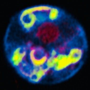
A color-enhanced image of a cell experiencing “stress” caused by alpha-synuclein expression, a protein associated with Lewy body dementias. The yellow color represents abnormal alpha-synuclein membranes.
Research Interests: The focus of my lab’s research has evolved from considering mechanisms that preserve chromosome stability and how they become compromised in aneuploid cancers to address a broader question about how cancer cells tolerate too many chromosomes:Our current focus is on identifying the stressors and pathways that lead to programmatic changes that allow disease states to persist. The observations driving this question are manifold but are nicely exemplified in hyperploid cancer cells that grow and divide in the presence of proteostatic and genomic stressors that are elevated due to too many chromosomes. In normal cells, such stressors would suppress cell proliferation and reduce cell viability. Yet in cancer cells proliferate and survive with these pathways chronically activated. Our work has identified a series of autophagy pathways connected to nuclear and endoplasmic reticulum homeostasis. These same pathways intersect with the cell stress associated with neurodegenerative disease models. Our work now is to understand how such pathways respond to acute cell stress and are altered in the context of “chronic cell stress”.
In posts under “Our Shared Biology”, I will share small stories about the cool things cells do and how it connects to larger questions about our shared biology, questions that we might be compelled to discuss as scientifically literate citizens. I know I’m not the only one who thinks about these issues or has great ideas about them, so please reach out and share. I’d love to hear from you! (kbkaplan@ucdavis.edu)
Our shared cell biology: I study cells. When I meet someone who asks me what I do, I often struggle to find an answer that feels adequate. Saying “I study cells” tends to end conversations pretty fast.So, instead I say that I study the cellular basis for disease; you know, like how normal cells become cancer cells. This is true enough and the mention of cancer, which touches so many, can start important conversations. Still, I can’t help feeling afterward that I’ve sold myself — and cell biology more generally — a bit short.There’s a more honest and “preachy” answer to the question of “what do you do?” that percolates inside my head but never quite makes it way out. It might sound something like this: “I am a cell biologist. I study cells because knowing how they work inevitably leads me (and us) to the much larger story of who we are, where we came from and (maybe) where we are going. To tell this story, I try to understand how cells adapt to perturbations in order to maintain their identity, or fail to adapt in the case of disease. It is in understanding and ultimately connecting how cellular machines make cells so robust across the diversity of life on our planet that our shared biology can be slowly revealed, and we can begin to appreciate the enormous impact we humans have on this narrative. This is what I do.”
If that sermon didn’t make the point clearly enough, I can also offer up countless examples of our beautiful cell biology. My favorite this week? Did you know that blast-fungal pathogens — the bane of rice farmers across the world — use autophagy and septins to create a high pressure cellular “gun” that can penetrate the cell wall of a leaf? Crazy biology, right? The really crazy thing is that we use these same cellular machines in ways that might be mechanistically verrrrry similar. Cool, huh?
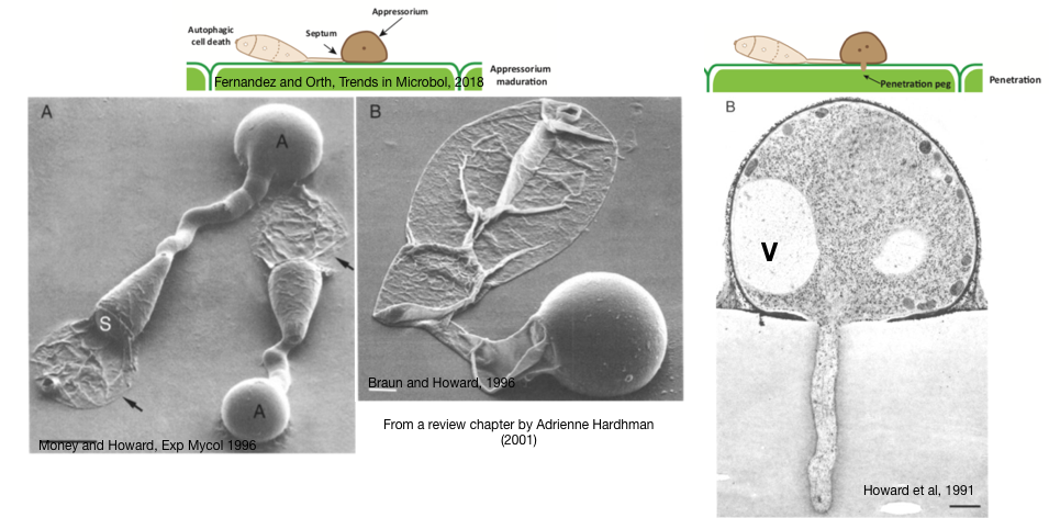
Blast Fungal Pathogens (for plants): The images below are scanning electron micrographs of one of these blast-fungal pathogens (Magnaporthe grisea, aka Magnaporthe Oryzea), This critter can grow as single cells, but under the right conditions comes together to make a three cell blast, or conidium. From the image below, you could be forgiven for thinking that a major part of the conidium has been “deflated”, because you’d be on the right track! The “stuff” inside the two “deflated” cells have in fact been degraded by a process called, autophagy (cell self-eating), where the contents of the cell are “shipped” off to the degradative organelle (i.e., vacuole or lysosome). In the middle image, you now see that the “germ tube” has also been deflated but the final area, the appressorium (A), does the opposite; it swells or inflates. In fact, the movement of all the degraded contents from the deflated cells of the conidium act to create pressure, which is funneled through to the appressorium and finally to a “penetration peg” (see the cartoons on top and the right hand image) that channels the turgor pressure (up to 8.0 MPa; 40X that of a car tire) to the growing peg in order to breakthrough the plant leaf and allow this alien-invader fungus to invade and spread INTO the plant cell. Penetration and invasion — yikes (think Alien, the movie)!
Electron micrographs of M. Oryzae, a fungal plant pathogen.
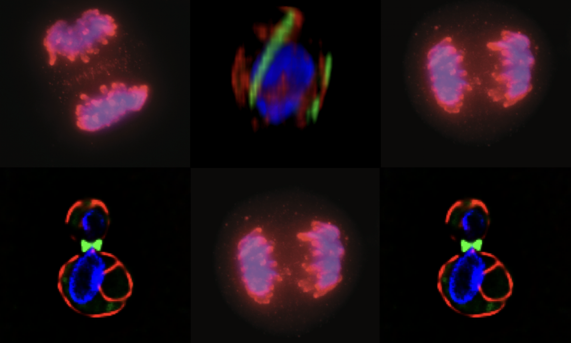
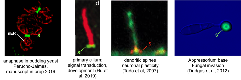
So what does this “Alien-like” biology have to do with our human-centric world view? Well, for one this blast-fungus is a major crop disease in rice and cereals, thus impacts the production of global food supply (not to mention having the potential to be a “weaponizable” biological weapon). We are also connected to this critter through the evolutionarily conserved cellular machinery that responds to environmental cues to organize the conidium into the invasion machine you see in the images. As the three cell conidium differentiates on the surface of the plant leaf, it begins to organize itself into distinct cellular regions by way of an understudied set of filaments called “septins”. Septins were first identified in the model organism budding yeast (S. Cerevisiae) where they were implicated in cell division. Since then, septins have been more broadly recognized to function in a diverse array of cell types in mammals. Septins are believed to be involved in forming physical barriers that regulate the diffusion of membrane proteins and contribute to the organization of such human-centric structures as primary cilia and dendritic spines, involved in key developmental signals and long-term potentiation in neurons, respectively (see example images below where “S” denotes septin structures). Therules that guide septin assembly in these circumstances remain unclear. Could a plant pathogen help us make sense of what is going on in our brains or during human development?
Examples of septin (see the “S”) filaments in fungi and mammalian cells. “nER” marks the nuclear endoplasmic reticulum.
The answer to that question is “to be determined”, but the key idea here is that it SHOULD be determined. Fungal genetics has already provided some important clues as to the signals that are important for septin organization during appressorium development. The proteins that control appessorium development are recognizable to students of human biology as parts of canonical signaling modules (i.e., trimeric G protein-coupled receptors, Ras and MAP kinase cascades, and transcription factors) that control everything from the flight-fight response to immune function in normal and cancer cells. Figuring out how these cell signal modules regulate septins in fungal plant pathogens will undoubtedly tell us something about how these same modules work in our cells. Yet, making such a case to the National Institutes of Health to study biology in model organisms, much less in a plant pathogen, is more and more an excuse to not fund a grant as it doesn’t make it over the human relevance bar. I think this overly human-centric view on biology hurts our chances to truly understand the complexity of our own biology, a biology that is part of a 4 billion year story of life on Earth that has by random chance allows us to thrive.More generally, human-centric constructs have not generally served us or our planet well. If our society solely invests in biology that leads to the next pharmaceutical, we will be missing out on important biology that has truly transformative potential. I join a growing set of voices that advocate for bringing the modern tools of cell biology (molecular genetics, biochemistry and live cell imaging) to understanding the diverse manifestations of life on our planet as a window to truly see how our shared biology fits into the narrative of who we are and where we are going. What do you think? Email me and let me know.
For more on how blast-fungal pathogens work and how they are connected to our conserved cell biology, check out these cool articles:
Van Ngo, H. & Mostowy, S. Role of septins in microbial infection. Journal of Cell Science132, jcs226266 (2019).
Hardham, A. R. in Biology of the Fungal Cell10, 91–123 (Springer, Berlin, Heidelberg, 2001)
Learning by doing is one of the most important and yet challenging goals to translate into the undergraduate experience. The act of “doing” allows students the chance to apply what they’ve learned in coursework to real world scenarios, to solve problems using critical thinking skills, and to reflect on how we understand the complex biology relevant to all of us. The goal of our upper division lab courses in MCB is to provide students access to learning by doing, albeit in a compressed 10-week format. I am seeking students interested in helping design new modules for the advanced cell biology lab course (MCB140L). Students will gain experience designing advanced curriculum and translating complex biological concepts and experimental approaches into practical modules. You can learn more about student projects here. Contact me (kbkaplan@ucdavis.edu) if you’re interested in participating (independent study units available).
Big congrats to all the undergraduate thesis students for 2019!
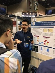
Jonathan Do, an Environmental Toxicology Major and(a McNair’s Scholar, joined the lab this year to explore how genome checkpoint pathways connect to our replication stress induced nucleophagy. Jonathan’s talents being a chemistry tutor are on full show here as he explains his thesis project to the rapt audience.
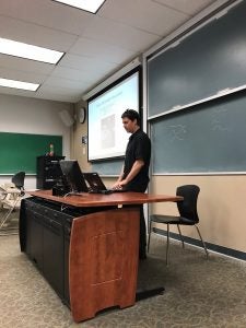
Mark Williams, a double Cell Biology and Physics major battled through computer issues to present his findings on how micronuclei, a major source of genome instability in tumor cells, might be suppressed by autophagy.
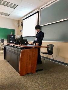
Mackenzie Noon, a Genetics major, presented his work on assessing how replication stress induced nucleophagy impacts the size of the rDNA array. Mackenzie has managed to develop a qPCR assay for measuring this chromosome array and is ready test his hypothesis.
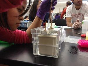
Ariana Cisneros, a Cell Biology major, presented her work tracking autophagy membranes in wild type and septin mutant cells. Ariana has spent a lot of time staring at “spots” in time-lapse images, so it’s no surprise that she’s in need of a cold beer after her talk (and I forgot to get a photo of her actual talk!!).
In January, we hosted Ryoya, a Ph.D student from the Nara Institute of Technology in Japan join us for a month long visit. Ryoya is from the Laboratory of Applied Stress and Microbiology at Nara, and is supervised by Dr. Takagi.
Ryoya teaching us about how to drink “brown wine”!
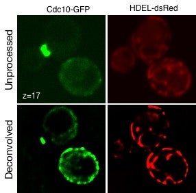
Ryoya helped us apply deconvolution algorithm to our spinning disc confocal data on septins. He learned how to use our experimentally determined point spread function data to reduce background, increase intensity and overall increase resolution of septin structures as observed below. Thanks for your hard work Ryoya and come back and visit any time.